Kidney stones constitute one of the commonest diseases in our country and pain due to kidney stones is known as worse than that of labour pain. In India, approximately 5 -7 million patients suffer from stone disease and at least 1/1000 of Indian population needs hospitalization due to kidney stone disease.
The kidney stone disease is widespread particularly in countries with dry, hot climate. These "stone belt regions" of the world are located in countries of Middle East – Dubai, Sharjah, Qatar, Muscat, Abu Dhabi, Saudi Arabia, North Africa, the Mediterranean Regions, North Western state of India and Southern State of USA and areas around the great lakes.
In India, the "stone belt" occupies parts of Maharashtra, Gujarat, Punjab, Haryana, Delhi and Rajasthan. In these regions, the disease is so prevalent that most of the members of a family will suffer from kidney stones sometime in their lives. Removal of Kidney, Ureteric and Bladder stone procedure forms one of the commonest procedures in Urology department of the hospitals in these regions.
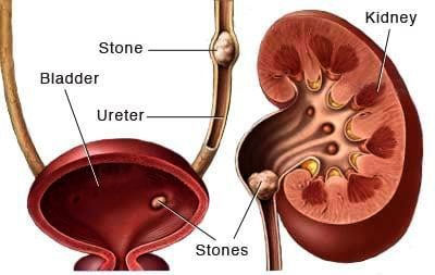
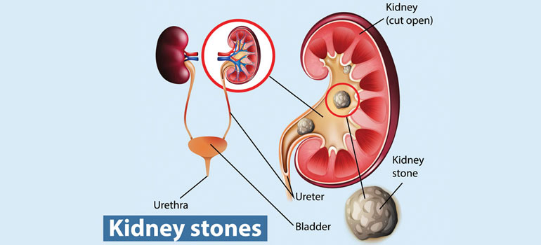
There are various types of urinary stones, but the most common ones are
In a rapid recurrent stone former – The metabolic activity for stone formation can be assessed by following investigations for the prevention of stone formation:
It has been said that "once a kidney stone former, always a kidney stone former". Once a kidney stone has been diagnosed, the choice is between expectant treatment and more aggressive forms of treatment, such as transurethral, percutaneous, or open surgeries or extra corporeal modalities. Although some kidney stones may pass spontaneously and unless complicating conditions arise, surgical intervention may not be necessary. Thus, identification of kidney stones that are likely to pass is of utmost importance.
The primary decision is whether to apply surgical treatment or wait. Removal of kidney stones by any methodology is necessary when there is evidence of:
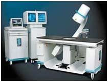
Extra Corporeal Shock wave Lithotripsy (ESWL) - Extra Corporeal Shock Wave Lithotripsy (ESWL) is a preferred mode of treatment for kidney stones upto 1.5 cm in size. An IVU is done prior to ESWL treatment to confirm the open passage from kidney to bladder for the finer fragment to pass out after a successful ESWL treatment. ESWL machine uses highly focussed sound wave projected from outside the body to break kidney stones. The stone is generally reduced to sand like particles which subsequently passes out in the urine. More than 1.5 cm to 2 cm stones generally requires more than one or two ESWL treatments.
The primary decision is whether to apply surgical treatment or wait. Removal of kidney stones by any methodology is necessary when there is evidence of:
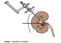
Percutaneous Nephrolithotripsy (PCNL) – Synonyms - Tunnel Surgery, Key Hole surgery for Kidney Stones
PCNL treatment is for a larger stone which are not indicated by ESWL. This procedure is generally done under general anaesthesia, spinal and /or epidural anaesthesia. In this technique the stone is removed by making a small tunnel into the kidney from the back. A fine needle is used to puncture the renal collecting system with the aid of X-ray and/or Ultrasonography, and a guide wire is led into the kidney through the needle. This tract is dilated over the guide wire and a Nephroscope (kidney telescope) is inserted into the pelvis of the kidney. The stones are visualized, fragmented using Swiss Lithoclast and extracted using fine forceps, allowing the kidney to become free of stones at the end of the operation, in the vast majority patients.
This Percutaneous Nephrolithotomy (PCNL) technique is used to treat kidney stones of: